


Euduboscquella anisospora

Diagnosis
Doubtful species.
Diagnosis_Genus: Euduboscquella Coats & Bachvaroff 2012. Euduboscquellidae with trophont episome as disc-shaped shield bordered by a perinematic ring. Lamina pharyngea extending from perinematic ring into trophont cytoplasm. Food vacuole formed as trophont emerges from host giving rise to extracellular tomont. Multiple spore morphotypes possible, including dinokont and non-dinokont cells. Individual infections producing only one type of spore.
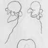
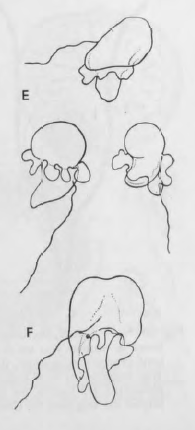
Diagnosis_Species: Trophont morphology/development: Similar to E. aspida but with several furrows that nearly encircle the body; undefined size
Sporogenesis: Not well described.
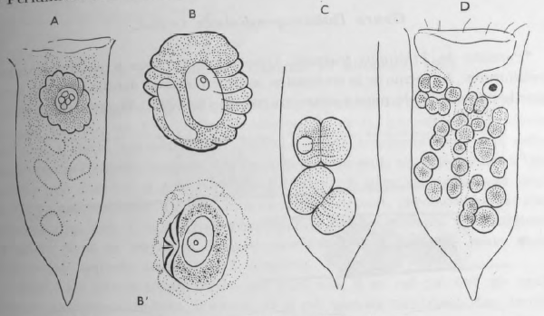
Spore morphology: Microspores 12.5 µm; macrospores 20 µm long; spores similar in shape with a globular episome and a narrower pointed hyposome; two flagella, one trailing and one transverse with large mastigonemes. Typical dinospores.
Body_macrospores_length: 20 µm
Body_microspores_length: 12.5 µm
Etymology
Genus name is derived from the Greek eu- (= well, normal) and the genus name Duboscquella.
Anisospora: typical dinospores (Syndiniales-like).
Type species
The type species of the genus is E. crenulata.
Type illustration / Type locality / Type specimen
Type host: Favella sp.
Ecology
Substrate_trophont: endozoic (endoparasite)
Substrate_tomont: extracellular
Substrate_spores: planktonic
Salinity: marine
Salinity: brackish
Salinity: variable
Life cycle
See the live cycle of E. aspida.
Phases_alternance: haplontic
Generation: <1 month
Reproduction_mode: asexual
Symbiont: horizontal_ingestion
