


Pelagococcus subviridis
Diagnosis
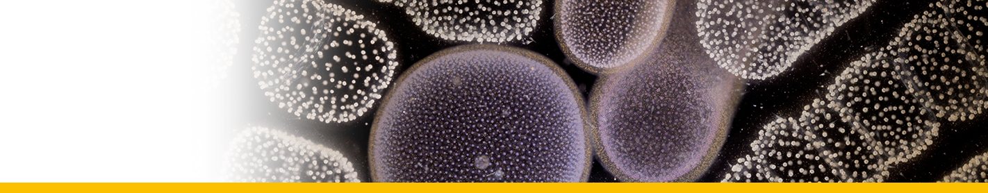
Cells pale green, spherroidal to ellipsoidal, sometimes having swollen protuberances. Cells occuring singly or weakly held together in irregular chains that may branch forming irregular colonies. Cell size ranging from 2.5 to 5.5 micrometers diam. Flagellate stages not present. Cells enclosed by a three layered cell wall having diffuse fibrillar material on the outer surface (possibly mucilage). Cultured cells adhere strongly to surface of glass container. Protoplasm consisting of a single, relatively large nucleus, a single chloroplast, one or possibly several mitochondria, a single dictyosome, endoplasmic reticulum and peripheral oblong bodies. Granular Polysaccharide reserve material not present. Chloroplast pigments are chlorophylls a, c diatoxanthin, diadinoxanthin, fucoxanthin like pigments, and carotene.
Type species
This is the type species (holotype) of the genus Pelagococcus.
Original description
Reference(s)
Observation site(s)
HOSTS
| Association with... | Region origin | Name of site | In reference... |
|---|---|---|---|
| Amphisolenia bidentata |
(2013) Using Nuclear-encoded LSU and SSU rDNA Sequences to Identify the Eukaryotic Endosymbiont in Amphisolenia bidentata (Dinophyceae). Protist 164:411 - 422. doi: 10.1016/j.protis.2012.10.001 |
