


Perkinsus marinus
Diagnosis
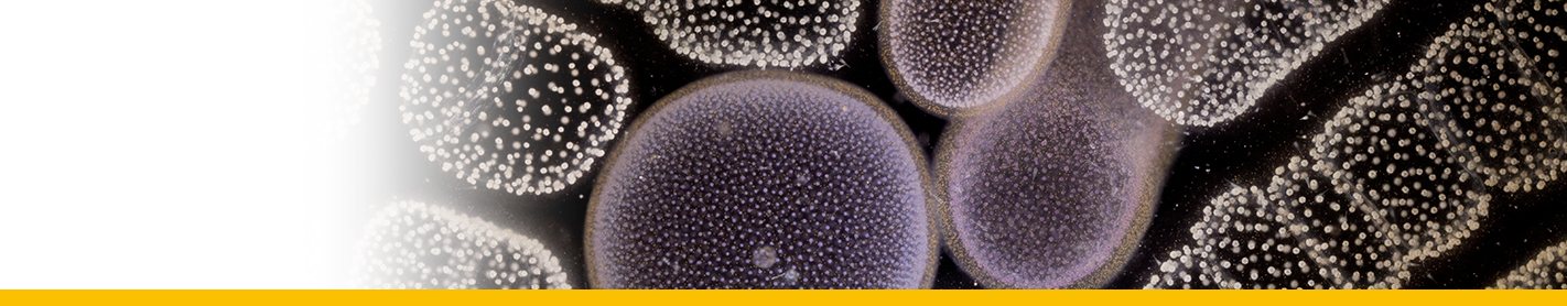
Diagnosis_Species: Studies of numerous stained sections and fresh preparationsshow that the most characeristic stage is a nearlyspherical spore, 3 to 10 µm in diameter ; themajority are 5 to 7 µm. Characteristically the cell contains a very large, slightly eccentric vacuole containing a large polymorphic refringent inclusion body, hereafter referred to as a vacuoplast. The cytoplasm of the cell forms a rather thin peripheral layer of alveolar nature,which is thicker on the side containing the nucleus. Occasionally the cytoplasm presents a slightly fibrous appearance and often contains aggregations of deep staining granules which are identical in staining reaction to the vacuoplast. The nucleus in the spore stage consists of a compact endosome surrounded by a clear zone usually free of chromatin material. The entire nucleus is ordinarily oval, the membrane often indistinct. The vacuoplast, when well formed, stains in shades of gray to black with Heidenhain's iron hematoxylin and stains a very light rose or diffuse pink with Delafield's hematoxylin and eosin. Because the vacuoplast stains heavily at the surface and less deeply internally, it often has the appearance of being hollow. Morphologically it may be a nearly prefect sphere, a lobular body, branched, or beaded. Often it is represented by several separate bodies or is broken into a large number of small granules, many of which may be distributed in small subvacuoles of the cytoplasmic layer. Occasionally binucleate stages are seen , the significance of which is not yet clear. Developmental stages of the spores usually have a vesicular type of nucleus, with some distributed chromatin and a less definite vacuole and vacuoplast. In reproduction, segmentation of the nucleus occurs with subsequent distribution of the daughter nuclei, followed by condensations of cytoplasm around each, and this in turn is followed by cytoplasmic cleavage and final liberation of sporelike cells by rupture of the thin containing membrane. The number of such cells produced is indeterminate, but occasionally as many as thirty may result from division of a single mother cell. Their size at liberation varies greatly. There are indications that endogenous budding occurs by a process of successive delaminations, producing spores of unequal size in an enveloping membrane formed from the parent cell. Any tissue of the host may be infected, common localizations being in the intestinal epithelium, adductor muscle, gills, mantle, and heart, or in cases of heavy infestation all tissues may be invaded. Development of the parasites is often intracellular in phagocytic or connective tissue cells.
Body_spores: 3-10 µm
Pigment: No
Toxicity: No
Allelopathy: No
Bloom: No (please provide a reference if yes)
Type illustration / Type locality / Type specimen
Type locality: Sugar House Bend in the southern part of Barataria Bay, Jefferson Parish, Louisiana.
Type material: Slides in the collections of the authors.
Type Host: Crassostrea virginica, the commercial oyster of the Atlantic and Gulf coasts.
