


Amoebophrya sticholonchae


Diagnosis
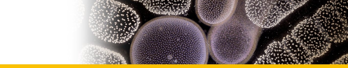
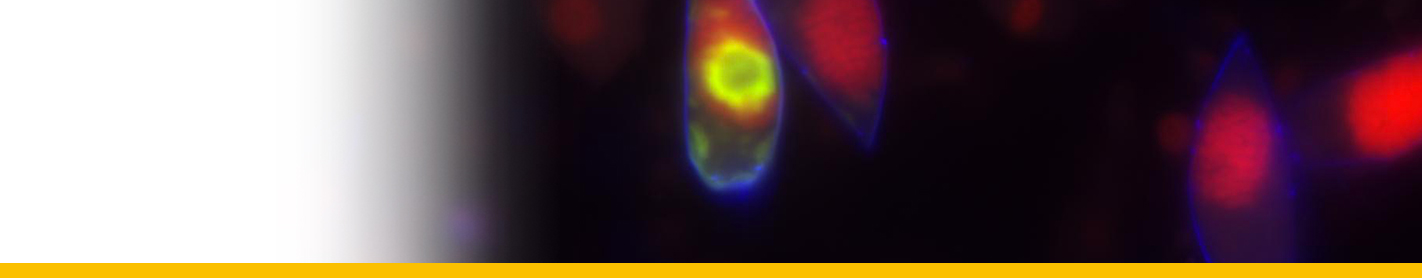
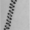
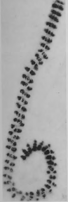
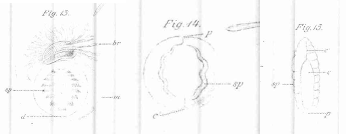
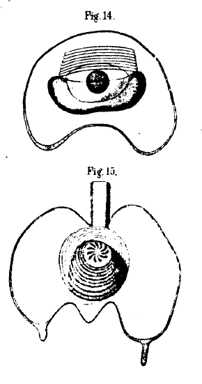
Diagnosis_Genus: Amoebophrya Koeppen 1984. Intracellular parasites. Smallest trophonts observed have 3 µm long, a gymnodiniales form, with a large hypocone and a small rudimentary epicone. The hypocone broadens at its base and forms a cup-shaped circumvallation, turning in spiral. The hypocone starts to cover the epicone (in other word, the epicone becomes progressively invaginated into the hypocone, forming a cavity, the mastigocoel, which is a characteristic of the genus Amoebophrya. This mastigocoel remains open by a small aperture at the apex of the trophont. Sporogenesis starts rapidly during this transformation. The inner surface of the mastigocoel is furnished with helically coiled ridges as a result of a continuous lengthening of the previous dinospore girdle during the trophic growth. Along these ridges countless flagella are inserted. Flagella of future dinospores (the free-living stage of the parasite) are produced inside the mastigocoel. At the end of the intracellular maturation, flagella started to beat, and what was the epicone at the origin, directed through the aperture of the mastigocoel, moved forward. This evagination make the flagella outside the structure, which is now called the vermiform. This action breacks out the host membrane, releasing the vermiform outside. This vermiform is closed forward (at the epicone side), but open in the back. When release, this part forms a large digestive vacuole that contains what remains from the host. This vermiform considerably strechs out with time. The spore chain forming the vermiform rapidly separates to produce spores (gymnodinial shape), generally of two different sizes (macrospores and microspores). These spores have a large epicone and a reduced hypocone (the reverse of what is observed during the first stage of the trophont).
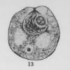
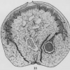
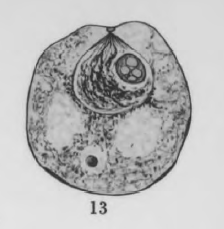
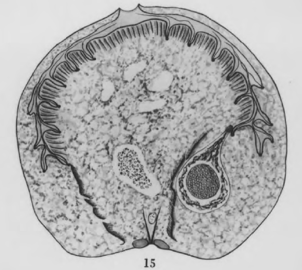
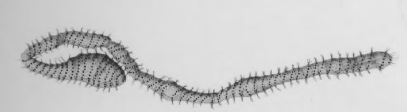
Diagnosis_species: Amoebophrya sticholonchae Koeppen 1894. The trophont develops outside of the central capsule of its host (Sticholonche), and could be considered as an ectoparasite. The trophont develops similarly to A. grassei, but could be either plurinucleate or having a single voluminous nucleus. A cytopharynx (see the description of Amoebophrya ceratii) is observed, but not well developped. The digestive vacuole of the vermiform includes the central capsule of its host, which persits for a long time before to be totally digested. Two type of spores (macro 7-8 µm long and microspores 2 µm long) are produced.
Body_spores_length: 7-8 µm (macrospores)
Body_spores_length: 2 µm (microspores)
Etymology
Amoebophrya: looks like to an amibe (Koeppen 1894).
sticholonchae: infects Sticholonche (zanclea), Koeppen 1894.
Type species
This is the type species of the genus.
Ecology
Substrate_spores: planktonic
Substrate_trophont: endozoic
Salinity: marine
pH: neutral
Feeding: parasitism (by phagotrophy)
Life cycle
Phases_alternance: haplontic
Generation: <1 month
Reproduction_mode: asexual
Symbiont: horizontal_active-penetration
Feeding behaviour
Mode of locomotion
Reference(s)
Observation site(s)
HOSTS
| Association with... | Region origin | Name of site | In reference... |
|---|---|---|---|
| Sticholonche zanclea |
(1894) Amoebophrya stycholonchae nov. gen. et sp. (corps spiral de Fol). Zoologischer Anzeiger 17:417-424. |
||
| Sticholonche zanclea | Villefranche-sur-mer |
(1883) Sur le Sticholonche zanclea et un nouvel ordre de Rhizopodes. Mem. Inst. Nat. Genevois 15:1-35. |
