


Chytriodinium affine
Diagnosis
Diagnosis_Genus: Ectoparasite of crustacean eggs.
Diagnosis_Species: C. affine. A dinospore attachs on the host egg (eggs have 60 to 90 µm in diameter, likely several host species are infected) and forms a cylindrical stalk. The stalk, which is the functional hypocone, penetrates the egg of the host and extended through the vitellus (the parasite nucleus remains outside). Upon reaching the embryo, the stalk enlarges at its base and forms a disk, develops rhizoids at its periphery and a feeding apparatus in its central part. This feeding apparatus consists in an ampulla having a large trophic vacuole that gradually absords the egg cytoplasm. The egg nucleus is also totally digested by the parasite, visibly by osmotrophy. The trophont nucleus has a synenergid appearance. The parasite then undergoes sporulation via palintomy, and produces 16 to 32 sporocytes. Successive synchronous divisions produce a long chain of communicating sporocytes having the same age, still surrounded by the external cuticle of the trophont. It seems that sporocytes are additionally retained by a common external membrane (Gomez et al. 2015). This sporocyte chain remains physically attached to the anchorage/feeding apparatus that is still functional and feds on the host during the first hours of the sporogenesis (observation until 24 hours). When all sporocytes are formed, the membrane containing the coiled chain of sporocytes detached from the empty egg, leaving the trophont external membrane that remained attached to the egg. The external cuticle still enclosing sporocytes ruptures when last generation of sporocytes are produced. Released into water, sporocytes develop flagella and undergo their final division. Mature dinospores possess two flagella, lack plastid, and are pyriform. They posess a typical dinocaryon. About 200 dinospores are produced per infected host. The dinospores of C. affine were able to infect new copepod eggs immediately after the last binary division.
Multi-infection was a common feature in Chytriodinium (Gomez et al. 2015), although no more than two trophonts developed successfully.
Body_spores_length: 9 µm
Body_spores_wide: 6 µm
Body_trophont: 20 µm
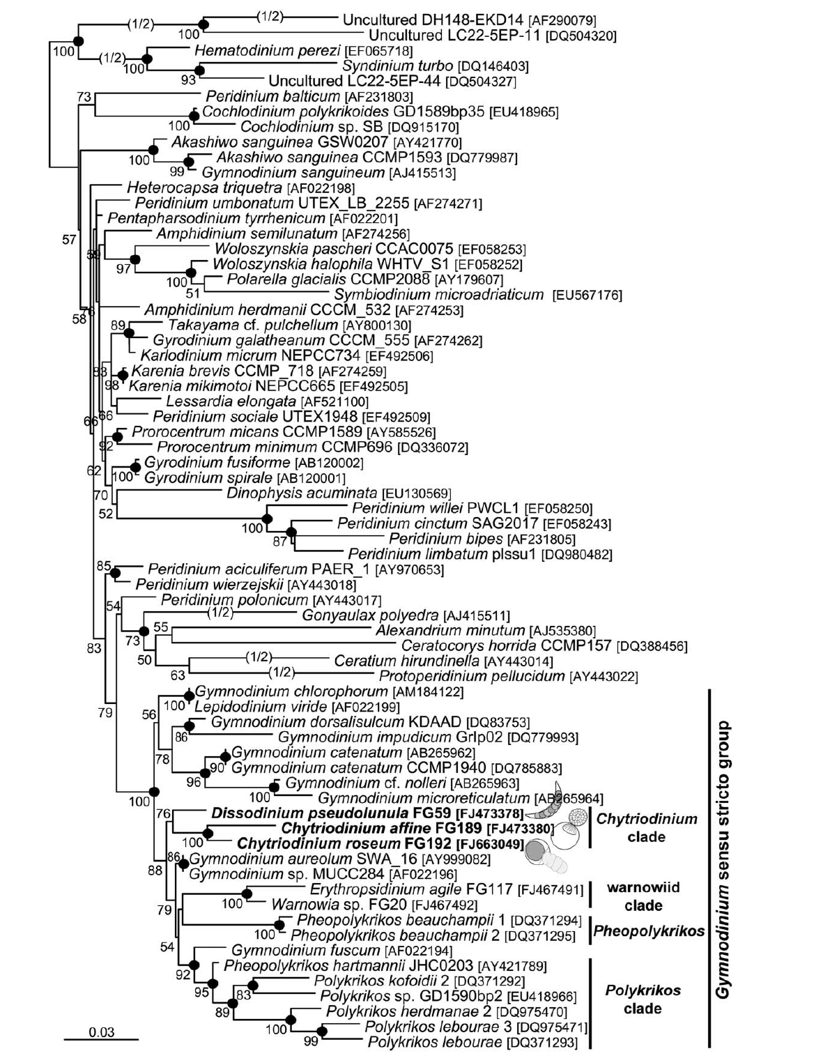
Sequence_SSU: FJ473380
Type species
The type species for the genus Chytriodinium roseum.
Type illustration / Type locality / Type specimen
Type host: copepod eggs
Ecology
Substrate: epizoic
Salinity: marine
Life cycle
Generation: <1 month
Reproduction_mode: asexual
Symbiont: horizontal
Feeding behaviour
Mode of locomotion
Reference(s)
Attached phylogeny
Observation site(s)
HOSTS
| Association with... | Region origin | Name of site | In reference... |
|---|---|---|---|
| Copepoda | Naples |
(1906) Beiträge zur Kenntnis des perininen. Mitt. aus der Zoll. St. zu Neapel XVIII:1-45. |
|
| Copepoda | Station Marine d'Endoume |
(2009) Life cycle and molecular phylogeny of the dinoflagellates Chytriodinium and Dissodinium, ectoparasites of copepod eggs. European Journal of Protistology 45:260 - 270. doi: 10.1016/j.ejop.2009.05.004 |
|
| Copepoda | Villefranche-sur-mer |
(1968) Cytologie et cycle évolutif des Chytriodinium (Chatton). Protistologia T. IV:249-262. |
