


Amoebophrya rosei


Diagnosis
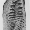
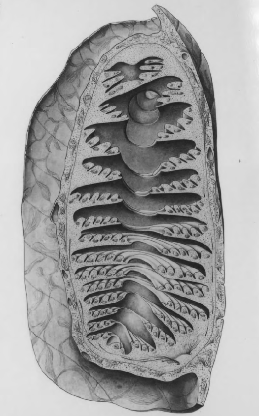
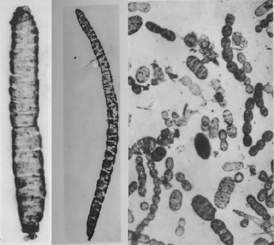
Diagnosis_Genus: Amoebophrya Koeppen 1984. Intracellular parasites. Smallest trophonts observed have 3 µm long, a gymnodiniales form, with a large hypocone and a small rudimentary epicone. The hypocone broadens at its base and forms a cup-shaped circumvallation, turning in spiral. The hypocone starts to cover the epicone (in other word, the epicone becomes progressively invaginated into the hypocone, forming a cavity, the mastigocoel, which is a characteristic of the genus Amoebophrya. This mastigocoel remains open by a small aperture at the apex of the trophont. Sporogenesis starts rapidly during this transformation. The inner surface of the mastigocoel is furnished with helically coiled ridges as a result of a continuous lengthening of the previous dinospore girdle during the trophic growth. Along these ridges countless flagella are inserted. Flagella of future dinospores (the free-living stage of the parasite) are produced inside the mastigocoel. At the end of the intracellular maturation, flagella started to beat, and what was the epicone at the origin, directed through the aperture of the mastigocoel, moved forward. This evagination make the flagella outside the structure, which is now called the vermiform. This action breacks out the host membrane, releasing the vermiform outside. This vermiform is closed forward (at the epicone side), but open in the back. When release, this part forms a large digestive vacuole that contains what remains from the host. This vermiform considerably strechs out with time. The spore chain forming the vermiform rapidly separates to produce spores (gymnodinial shape), generally of two different sizes (macrospores and microspores). These spores have a large epicone and a reduced hypocone (the reverse of what is observed during the first stage of the trophont).
Diagnosis_species: Hyperparasite of apostome parasites of Siphonophores (Abylopsis tetragona, Rose 1937), and Chaetognathes (inside the posterior intestin of Sagitta). Located first closely related to the lipidic vacuole of the ciliate, the young trophont then develops close to the nucleus. Compared to other Amoebophrya species, A. rosei has a rather reduced mastigocoel. First nuclear mitotic divisions are enclosed inside the mother nuclear membrane. After more than 200 nuclear divisions, this external membrane disappears. Mature trophont has 150 µm long and 70 µm width. Cytopharynx (as described for Amoebophrya ceratii) is absent. The maturation end only when the apostome is released from the Siphonophore.
Body_trophont_length: 150 µm
Body_trophont_Width: 70 µm
Type species
The type species (lectotype) of the genus Amoebophrya is Amoebophrya acanthometrae Borgert 1897
Type illustration / Type locality / Type specimen
Type_host: Hyperparasite of apostome parasites of Siphonophores (Abylopsis tetragona, Rose 1937), and Chaetognathes (inside the posterior intestin of Sagitta)
Ecology
Substrate_spores: planktonic
Substrate_trophont: endozoic
Sociability_spores: solitary
Sociability-trophont: solitary
Salinity: marine
pH: neutral
Feeding: hyperparasitism (by phagotrophy)
Life cycle
Phases_alternance: haplontic
Generation: <1 month
Reproduction_mode: asexual
Symbiont: horizontal_active-penetration
Feeding behaviour
Mode of locomotion
Reference(s)
Observation site(s)
HOSTS
| Association with... | Region origin | Name of site | In reference... |
|---|---|---|---|
| Apostome from the Siphonophore Abylopsis tetragona |
(1964) Contribution à l’étude des péridiniens parasites. Cytologie, cycles évolutifs. Ann. Sci. Nat. Zool. 6:1-158. |
